Where Is Your Ciliary Ganglion
These paired ganglia supply all parasympathetic innervation to the head and neck. -Connected to nasociliary N.
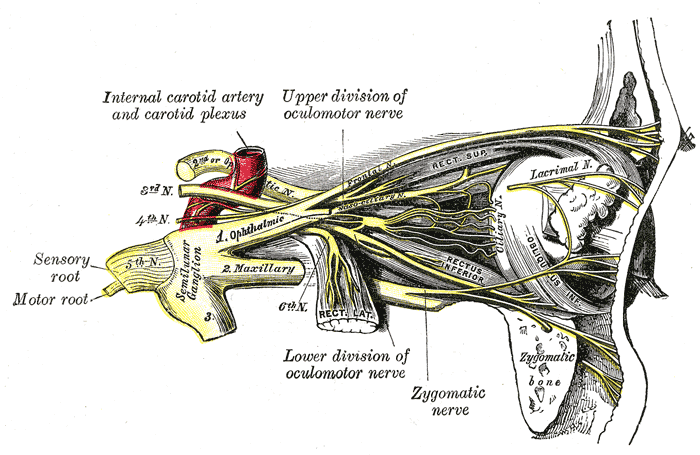
It is situated at the back part of the orbit in some loose fat between the optic nerve and the Rectus lateralis muscle lying generally on the lateral side of the ophthalmic artery.
Where is your ciliary ganglion. It is a relay station for fibers supplying sphincter pupillae and ciliaris muscle. It is approximately 15-20 cm 15-20 mm posterior to the globe and 10 cm 10 mm anterior to the Annulus of Zinn and the superior orbital fissure. It receives parasympathetic fibres from the oculomotor nerve.
Ciliary ganglion spincter pupillae ciliary muscle pterygopalatine ganglion lacrimal gland glands of nasal cavity submandibular ganglion submandibular and sublingual glands and otic ganglion parotid gland. The ciliary ganglion lies temporal to the ophthalmic artery inbetween the lateral rectus and optic nerve. There may be one or more accessory ganglia.
A dorsal root ganglion or spinal ganglion is a nodule on a dorsal root of the spine that contains the cell bodies of nerve cells neurons that carry signals from the sensory organs towards the appropriate integration center. It receives parasympathetic fibers from the oculomotor nerve. 52 Pathways Distribution 1 Ciliary ganglion -lies in the orbit lateral to the optic N.
The ciliary ganglion is important when considering CN V 1 specifically the nasociliary divisionThe ganglion is formed by cell bodies of postganglionic parasympathetic neurons and is located toward the apex of the orbit between the optic nerve and the lateral rectus muscle Figure 224. -Edinger-Westphal nucleus in the upper part of midbrain. The ciliary ganglion ophthalmic or lenticular ganglion is a small sympathetic ganglion of a reddish-gray color and about the size of a pins head.
NER Module 75 Summary of the Parasympathetic Ganglia. The ganglion is found on the anterior surface of the petrous part of the temporal bone in a dural pouch known as Meckels cave. Both of these muscles are involuntary since they are controlled by the parasympathetic.
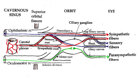
Preganglionic parasympathetic pupillomotor fibers cell bodies in Edinger-Westphal. This ganglion contains the cell bodies of the postganglionic pupilloconstrictor neurons which innervate the sphincter muscle of the iris. It is one of four parasympathetic ganglia of the head and neck.
The ciliary ganglion lies behind the eye. Eg an accessory ganglion has been found on the medial side of the optic nerve. Gross anatomy smallest of the ganglia 2 mm in size located posterolaterally in the intraconal spa.
Medical Definition of ciliary ganglion. The ganglion and its site Nucleus and its site Fig. Branch to inferior oblique muscle.
It is 12 mm in diameter and in humans contains approximately 2500 neurons. It is situated anteriorly to the superior orbital fissure between the lateral rectus muscle and the optic nerve. The ciliary ganglion is one of four parasympathetic ganglia of the head and neck.
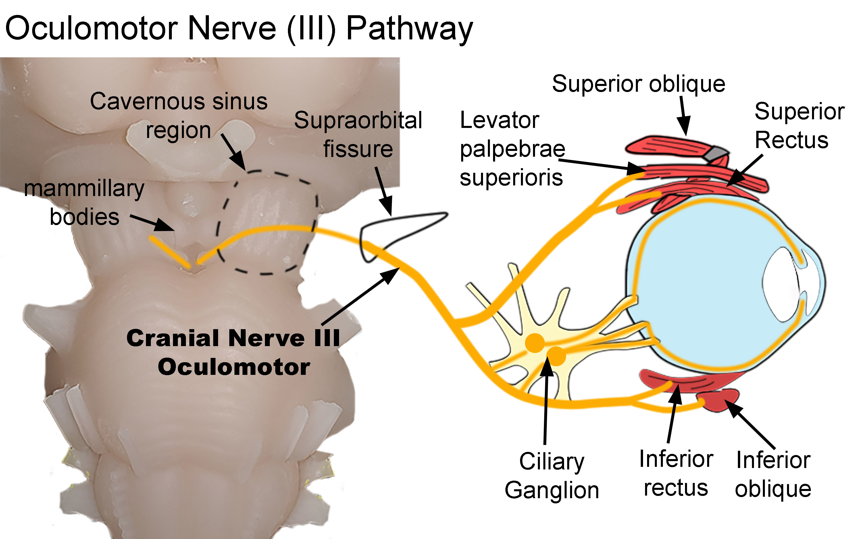
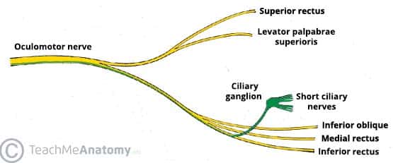
The ciliary ganglion has been found perforated by a ciliary artery. The ciliary ganglion receives preganglionic fibers via the oculomotor nerve cranial nerve III that originate from the Edinger-Westphal nucleus in the midbrain. The ciliary ganglion is one of four parasympathetic ganglia of the head and neck.
There is a shred of existing evidence in the literature that ciliary muscle also receives innervation from the sympathetic fibers of the autonomic nervous system ANS. Nerves that carry signals towards the brain are known as afferent nerves. It is topographically connected to nasociliary nerve branch of ophthalmic division of trigeminal nerve It is functionally related to Oculomotor nerve.
It is a parasympathetic ganglion a type of autonomic ganglion. The ciliary ganglion is approximately 3 mm in size and located 23 mm posterior to the globe and lateral to the optic nerve. The trigeminal ganglion is the largest of the cranial nerve ganglia.
The exceptions are the four paired parasympathetic ganglia of the head and neck. The entering axons are arranged into three roots of the ciliary ganglion which join enter the posterior surface of the ganglion. The ganglion contains postganglionic parasympathetic neurons.
The ciliary ganglion lies within the lateral orbit between the optic nerve and lateral rectus muscle. It is approximately 1 cm in front of the annulus of Zinn. Seifert in Nerves and Nerve Injuries 2015 Ciliary Ganglion.
The ciliary ganglion is a parasympathetic ganglion located just behind the eye in the posterior orbitThree types of axons enter the ciliary ganglion but only the preganglionic parasympathetic axons synapse there. The ciliary ganglion is a bundle of nerve parasympathetic ganglion located just behind the eye in the posterior orbit. The ciliary ganglion is supplied by fibres from the Edinger-Westphal nucleus associated with the oculomotor nerve.
These neurons supply the pupillary sphincter muscle which constricts the pupil and the ciliary muscle which contracts to make the lens more convex. Inferior division of CN III. The ciliary ganglion is located within the bony orbit.
Nerves From the cavernous sinus. Ciliary Ganglion It is a Parasympathetic ganglion. A small autonomic ganglion on the nasociliary branch of the ophthalmic nerve receiving preganglionic fibers from the oculomotor nerve and sending postganglionic fibers to the ciliary muscle and to the sphincter pupillae called also lenticular ganglion Learn More about ciliary ganglion.
 Opt 114 3b Outer Tunic Flashcards Quizlet
 Pdf Parasympathetic Ganglia In The Head
Sensory Root Of Ciliary Ganglion Nasociliary Root Of Ciliary Ganglion Communicating Branch Of Nasociliary Nerve With Ciliary Ganglion
 Parasympathetic Innervation To The Head And Neck Anatomy Ganglia Teachmeanatomy
 Ciliary Ganglion Autonomic Control Of The Eye Anatomy Tutorial Youtube
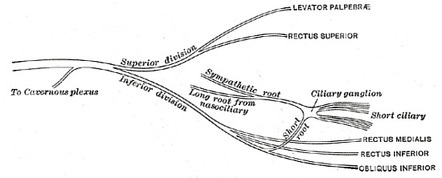 Ciliary Ganglion Radiology Reference Article Radiopaedia Org
 Ciliary Ganglion Location Roots And Branches Anatomy Qa
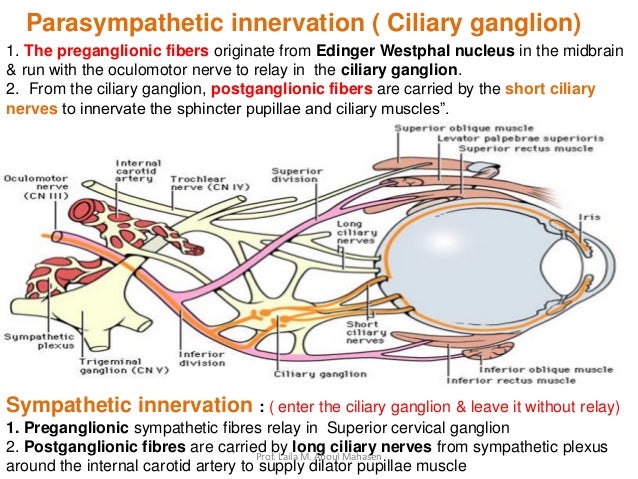 Prof Laila 2017 Kau Parasympathetic Ganglia
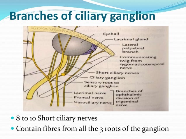 Parasympathetic Ganglia Of Head And Neck
 Anatomy Of Ciliary Ganglion Youtube
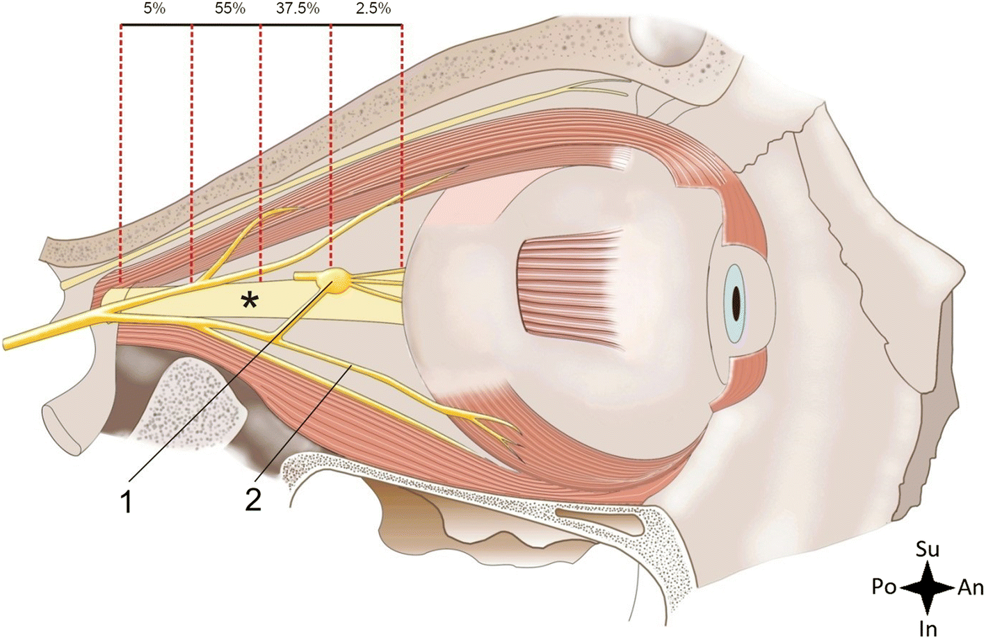 Figure 1 Anatomical Variations Of The Ciliary Ganglion With An Emphasis On The Location In The Orbit Springerlink
 Physiology Test 4 Nerve Functioning Flashcards Quizlet
:background_color(FFFFFF):format(jpeg)/images/article/en/parasympathetic-ganglia/LZ7Hpv9PHYoNFOFJbNc8XA_AjiKaS2VMPFebQpMxFPA_Parasympathetic_root_of_ciliary_ganglion_1.png) Parasympathetic Ganglia Anatomy And Function Kenhub
Parasympathetic Ganglia Anatomy And Function Kenhub
 Ciliary Ganglion Situation Relations Connections Branches Distribution Youtube
